Dental X-Rays

X-ray examination of teeth is an essential diagnostic procedure that allows specialists to determine the diagnosis of a patient’s diagnosis and the sequence of further actions. In treating many diseases, this is the first step on the road to recovery.
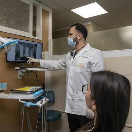
This survey is based on gamma radiation. X-rays pass through the patient’s organic tissues, resulting in an image on a special film or a computer screen.

X-ray images demonstrate the condition of bone tissue and make it possible to identify many negative changes. The doctor sees what is hidden from the naked eye and determines if the patient has tooth decay, gum disease, abscesses, or abnormal growths such as a tumor or cyst. In addition, X-rays allow you to determine the location and condition of the affected or unerupted teeth.

Types of images →

At Aesthetic Dental, dentists use a new generation of X-ray machines, which have a number of advantages – minimal radiation, quick examination, and obtaining results on the day of the investigation, which is essential for timely diagnosis.

Aesthetic Dental doctors receive information about the patient from several types of X-rays of the dentoalveolar system.

A sighting snapshot of the teeth shows the condition of one or more teeth and adjacent tissues. This helps the specialist to assess the condition of dentin, root canals, periodontium, and blood vessels.

A dental X-ray or orthopantomogram is a detailed flat image of the jaws, teeth, joints, and sinuses. Such a picture helps to detect carious areas, defects in fillings, impacted teeth, and cysts. And also draw up a correct plan for implantation, prosthetics, and treatment.

Occlusal scan, or X-ray bite, is often used in examining children and when it is impossible to obtain precise results using targeted radiography.

Computed tomography, as a research method, is the most informative. The digital format with the effect of 3D visualization displays the jaw projection in a three-dimensional version. The effect of radiation is negligible due to the short duration of the procedure.

During the operation of the tomograph, the picture is transmitted to the monitor screen. The Aesthetic Dental doctor immediately works on the diagnosis and treatment of the patient and records the result on digital media.

Deciphering the received data →

Aesthetic Dental doctors consider this procedure a reasonably informative diagnostic method to identify pathological processes. The picture informs doctors about this area’s inflammatory process, atrophy, neoplasm, and the initial signs of caries.

The essential step is interpreting the image obtained during the study. The tonality of the color allows you to accurately diagnose the condition of tissues – healthy, with pathologies, with tumors or cysts, inflamed or necrotic.

Healthy tissues of the gums and teeth in the picture are painted gray, and fillings and implants will be a characteristic white color. A tooth cyst and other neoplasms will be displayed as a dark area with precise contours with a rounded or oblong shape.

The use of X-rays with a contrast agent has indicated if it is necessary to assess the condition of the ducts of the salivary glands. This type of diagnosis allows you to identify some pathologies.

The practicality of conducting an X-ray also arises if dental treatment has already been carried out. Still, the dentist needs to ensure there are no complications and evaluate the quality of the procedure.

Bite correction is also necessarily accompanied by an X-ray at all stages of this process.

Harm, contraindications, and indications→

Questioning and examining the patient gives the doctor only part of the information about the natural state of the dentoalveolar system. To clarify the alleged diagnosis, in many cases, it is required to undergo an X-ray examination. But the very mention of it annoys some people. Is this procedure hazardous to health?

Exposure to X-rays, in one way or another, negatively affects the human body’s cells. The bottom line is only in the dosage of this radiation and the frequency of procedures.

The radiation dose for dental X-rays that the patient receives varies from 5 micro Sieverts to 700 micro Sieverts, depending on the image’s size and the X-ray machine’s model. And one procedure using a digital radiovisiograph is equivalent to 2 micro Sieverts.

To understand the above, we can compare these data with the average dose of natural background exposure in the environment received by a person per year. The average international dose is 2400 microsieverts per year.

By the standards, the maximum permissible annual radiation dose for diagnostic X-ray procedures is 1000 microsieverts for adults and 300-400 microsieverts for a child.

To obtain it, during the year, it is necessary to undergo 40 film orthopantomography to 500 targeted examinations of teeth using a radiovisiograph. Thus, dental X-rays for almost any appointment do not go beyond the permissible doses.

When choosing an X-ray, Aesthetic Dental doctors, to reduce the patient’s exposure, follow the international principle of ALARA (As Low As Reasonably Achievable) – using the lowest and most effective radiation dose. The dentist should recommend X-rays only to the extent necessary for optimal oral health.

The dose of radiation exposure during X-ray examination is minimal. But this procedure can be carried out with restrictions.

The main contraindications include pregnancy, problems with low immunity, mental disorders, and bleeding.

The question of X-rays in pregnant women is quite acute. This procedure is possible even in this case. The difficulty lies in the fact that it is impossible to determine the exact effect of radiation on the structure of the fetus, so there are certain limitations. A nursing woman should use digital methods and skip one or two feedings after the session.

At the Aesthetic Dental clinic, you can undergo high-precision dental diagnostics using a modern X-ray machine, which guarantees safety for the body.

At the Aesthetic Dental clinic, you can undergo high-precision dental diagnostics using a modern X-ray machine, which guarantees safety for the body.

Identification of pathologies is not the only indication for this study. X-rays of the tooth are prescribed for all types of endodontic and surgical treatment and for quality control of the procedures performed. Radiography is an essential diagnostic tool for the dentist. Early detection of the disease and timely treatment is the best way to keep your teeth healthy for life.

If the patient has doubts about the type and quantity of X-rays recommended by the dentist Aesthetic Dental, the patient will always be able to ask questions and get a qualified answer.

FAQ

A dental image allows you to clarify the diagnosis, assess the general condition and internal structure of the tooth, the condition of bone tissue and gums, and choose the right treatment tactics. To control the treatment, taking an X-ray of the teeth is necessary.

A sighting image of one tooth equals 2-3 micro Sieverts. You can take 300-500 X-rays per year. A panoramic image – all teeth – resembles 16-18 micro Sieverts. You can take 60-70 X-rays per year.

Orthopantomogram or panoramic image of the teeth and jaw – an image of all teeth, bone tissue of the upper and lower jaw, the structure of the maxillary sinus, and the temporomandibular joint.

A panoramic image is one of the most common methods for diagnosing pulpitis. The X-ray shows several signs indicating pulpitis. Pronounced darkening in the area of root canals or dental cavities suggests the presence of an inflammatory process.

Today, research facilities are being modernized, and their harm is minimized, so it is impossible to say which is more harmful, X-rays or computed tomography. However, X-rays are generally safer than CT, although less informative.